Researchers from the Universities of Münster and Potsdam developed a method for the quantitative bioimaging of mercury in fruit flies for investigating the uptake of different mercury species.
Background:
The heavy metal mercury has been known as a toxic element for centuries. From the mass poisoning events of Minamata and Iraq poison grain disaster it is well known that the toxicity is significantly depending on the chemical species being present. Although the toxicology of mercury is a highly investigated topic, much less is known about the transport, distribution and metabolism of mercury species in biological organisms.
It is well understood that mercury shows a high affinity to sulfhydryl groups, which are omnipresent in proteins serving as binding partners in adduct formation. For this reason, mercury accumulates in those parts of the organism rich in the amino acid cysteine. Further, it was shown that organomercury compounds such as methylmercury and ethylmercury are able to cross the blood-brain barrier when bound to particular amino acids acting as transporters. In order to study such processes in more detail, there is increasing interest to visualize the mercury distribution in relevant biological tissues. While laser ablation with inductively coupled plasma mass spectrometric detection (LA-ICP-MS) has been developed as a versatile tool for bioimaging purposes during recent years, quantification of mercury in LA-ICP-MS studies remains challenging. Major problems are analyte losses by volatilization and adsorption as well as matrix effects.
The new study:
German researchers from the Universities of Münster and Potsdam developed a method for the quantitative bioimaging of mercury in fruit flies used as a model organism. The use of this model organism allows for a high number of experiments with different mercury species and improved statistical conclusions for both practical and ethics reasons.
Wild type fruit flies (Drosophila melanogaster) were exposed to mercury through feeding- stuff contaminated with methylmercury, thimerosal or mercury chloride of various concentrations. Ten female and male parental animals were raised up in tubes containing the mercury spiked feed in incubators at 25 °C with artificially generated 12 h/12 h light rhythm. After 3 days of breeding and sufficient oviposition, the parental generation was removed. L3 state larvae were collected after 9 days and the adult F1 generation after 14 days of breeding.
For later bioimaging purpose the sacrificed flies and larvae were infiltrated in sucrose solution and embedded in gelatin. The samples were cut with a cryotome into sections having a thickness of 10 or 20 µm.
For external calibration, gelatin-based matrix-matched standards were prepared. Gelatin proved to be a suitable matrix as it is easy to handle and consists of proteins so that the laser ablation properties can be compared with the biological tissues of larvae or adult flies. Mercury volatilization losses were avoided by adding DMSA as complexing agent.
The analysis of samples and standards via LA-ICPMS was performed with a commercially available laser ablation system (LSX 213 G2+, CETAC Technologies, Omaha) coupled to a quadrupole based inductively coupled plasma mass spectrometer (Agilent 7500 ce, Santa Clara). Due to the known neurotoxicity of some mercury species, one of the main targets for further analysis was the spatial distribution of mercury in the brain and the selectivity of the blood-brain barrier.

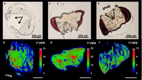
Fig. 2
(a) Microscopic and (b) LA-ICPMS mercury maps of a fruit fly larva fed with methylmercury chloride (0.2 µg Hg/g). The lobes of the brain are marked with arrows. (c) Microscopic and (d) LA-ICPMS image of the head of an adult fruit fly fed with methylmercury chloride (0.2 µg Hg/g). (e) Microscopic and (f) LA-ICPMS image of the head of an adult fruit fly fed with thimerosal (0.2 µg Hg/g). The
202Hg signal is used for data evaluation. Images were recorded with a laser spot diameter of 10 µm and a cutting thickness of 20 µm.
The LA-ICP-MS mercury maps for the larva clearly show a high enrichment of 10- 20 µg/g mercury in the brain, corresponding to a factor of 50 compared to the mercury concentration in feed. Thus, the expected transfer of methylmercury chloride over the blood-brain barrier can be confirmed.
The different accumulation behavior of methylmercury and ethylmercury could be observed in the brains of adult flies. For both, methylmercury chloride and thimerosal, mercury was detected within the whole head. Again, this indicates that the used organic mercury species are able to pass the blood-brain barrier. The enrichment of methylmercury in the brain is approximately 3 times higher than for thimerosal. The investigation of even higher feed concentrations of mercury(II) chloride also showed the absence of mercury in the flys head indicating that this inorganic species cannot cross the blood-brain barrier.
 The original studies

Ann-Christin Niehoff, Oliver Bolle Bauer, Sabrina Kröger, Stefanie Fingerhut, Jacqueline Schulz, Sören Meyer,
Michael Sperling, Astrid Jeibmann,
Tanja Schwerdtle,
Uwe Karst,
Quantitative Bioimaging to Investigate the Uptake of Mercury Species in Drosophila melanogaster, Anal. Chem., 87 (2015) 10392-10396.
DOI: 10.1021/acs.analchem.5b02500
A
nalytical instrumentation:
 Agilent ICP-MS 7500-CE
 Teledyne CETAC laser ablation system LSX-213 G2+
 Related Studies (newest first)

Tracy C. MacDonald, Malgorzata Korbas, Ashley K. James, Nicole J. Sylvain, Mark J. Hackett, Susan Nehzati, Patrick H. Krone, Graham N. George and Ingrid J. Pickering,
Interaction of mercury and selenium in the larval stage zebrafish vertebrate model, Metallomics, 7 (2015) 1247-12355.
DOI: 10.1039/c5mt00145e
M. Mela, F.F. Neto, S.R. Grötzner, I.S. Rabitto, D.F. Ventura, C. A. Oliveira Ribeiro,
Localization of inorganic and organic mercury in the liver and kidney of Cyprinus carpio by autometallography, J. Braz. Soc. Ecotoxicol., 7/2 (2012) 73-78.
DOI: 10.5132/jbse.2012.02.011
Benjamin D. Barst, Amanda K. Gevertz, Matthew M. Chumchal, James D. Smith, Thomas R. Rainwater, Paul E. Drevnick, Karista E. Hudelson, Aaron Hart, Guido F. Verbeck, Aaron P. Roberts,
Laser Ablation ICP-MS Co-Localization of Mercury and Immune Response in Fish, Environ. Sci. Technol., 45 (2011) 89828988.
DOIi: 10.1021/es201641x

Emiko Nakazawa, Tokutaka Ikemoto, Akiko Hokura, Yasuko Terada, Takashi Kunito, Shinsuke Tanabe, Izumi Nakai,
The presence of mercury selenide in various tissues of the striped dolphin: evidence from l-XRF-XRD and XAFS analyses, Metallomics, 3 (2011) 719725.
DOI: 10.1039/c0mt00106f
Dirce Pozebon, Valderi L. Dressler, Marcia Foster Mesko, Andreas Matusch and
J. Sabine Becker,
Bioimaging of metals in thin mouse brain section by laser ablation inductively coupled plasma mass spectrometry: novel online quantification strategy using aqueous standards, J. Anal. At. Spectrom., 2010, 25, 17391744.
DOI: 10.1039/c0ja00055h

Malgorzata Korbas, John L. ODonoghue, Gene E. Watson, Ingrid J. Pickering, Satya P. Singh, Gary J. Myers, Thomas W. Clarkson, Graham N. George,
The Chemical Nature of Mercury in Human Brain Following Poisoning or Environmental Exposure, ACS Chem. Neurosci., 1 (2010) 810818.
DOI: 10.1021/cn1000765
Maritana Mela, Sebastien Cambier, Nathalie Mesmer-Dudons, Alexia Legeay, Sonia Regina Grötzner, Ciro Alberto de Oliveira Ribeiro, Dora Fix Ventura, Jean-Charles Massabuau,
Methylmercury localization in Danio rerio retina after trophic and subchronic exposure: A basis for neurotoxicology, NeuroToxicology, 31 (2010) 448453.
DOI: 10.1016/j.neuro.2010.04.009

Malgorzata Korbas, Patrick H. Krone, Ingrid J. Pickering, Graham N. George,
Dynamic accumulation and redistribution of methylmercury in the lens of developing zebrafish embryos and larvae, J. Biol. Inorg. Chem., 15 (2010) 11371145.
DOI: 10.1007/s00775-010-0674-6
Richard R. Chapleau, Martin Sagermann,
Real-time in vivo imaging of mercury uptake in Caenorhabditis elegans through the foodchain, Toxicology 261 (2009) 136142.
DOI: 10.1016/j.tox.2009.05.005

Malgorzata Korbas, Scott R. Blechinger, Patrick H. Krone, Ingrid J. Pickering, Graham N. George,
Localizing organomercury uptake and accumulation in zebrafish larvae at the tissue and cellular level, Proc. Natl. Acad., Sci. USA, 105 (2008) 12108-12112.
DOI: 10.1073/pnas.0803147105
B. Inza, C. Rouleau, H. Tjälve, F. Ribeyre, P.G.C. Campbell, E. Pelletier, A. Boudou,
Fine-Scale Tissue Distribution of Cadmium, Inorganic Mercury, and Methylmercury in Nymphs of the Burrowing Mayfly Hexagenia rigida Studied by Whole-Body Autoradiography, Environ. Res. A, 85 (2001) 265-271.
DOI:10.1006/enrs.2000.4228
Erik Baatrup, Mogens Gissel Nielsen, Gorm Danscher,
Histochemical Demonstration of Two Mercury Pools in Trout Tissues: Mercury in Kidney and Liver after Mercuric Chloride Exposure, Ecotoxicol. Environ. Safety, 12 (1986) 267-282.
DOI: 10.1016/0147-6513(86)90018-7 Related EVISA Resources
 Brief summary: ICP-MS: A versatile detection system for speciation analysis
 Link Database: Toxicity of Organo-mercury compounds
  Link Database: Mercury exposure through the diet
  Link Database: Environmental cycling of methylmercury
 Link Database: Environmental cycling of inorganic mercury
 Link Database: Environmental pollution of methylmercury
 Link Database: Environmental pollution of inorganic mercury
 Link Database: Toxicity of mercury
  Link Database: Research projects related to organo-mercury compounds
 Link Database: All about thimerosal (thiomersal)
 Related EVISA News
 October 19, 2015: New mercury transporting protein in human blood identified
  September 7, 2014: New study finds relationship between organic mercury exposure from Thimerosal-containing vaccines and neurodevelopmental disorders
  December 29, 2013: A new study finds: Inorganic mercury stays in the brain for years if not decades
  November 9, 2013: Spatially resolved speciation analysis: A new technique based on laser ablation with simultaneous elemental and molecular mass spectrometry
  September 12, 2013: Scientists reveal how organic mercury can interfere with vision
  October 12, 2012: Prenatal mercury intake linked to ADHD
  June 19, 2012: Vaccine ingredient causes brain damage; some nutrients prevent it
  March 1, 2012: High levels of mercury in newborns likely from mothers eating contaminated fish
  August 15, 2011: New insights about interaction of thimerosal with blood components
  September 25, 2010: The European Chemical Agency (ECHA) calls for
comments on reports proposing restrictions on mercury and phenylmercury
 August 16, 2010: Methylmercury: What have we learned from Minamata Bay?
 September 24, 2009: Huge field experiment for assessing human ethylmercury risk starting in october
 July 15, 2009: New Study Finds: Thimerosal Induces Autism-like Neurotoxicity
 March 24, 2006: Mercury Containing Preservative Alters Immune Function
 February 11, 2005: New findings about Thimerosal Neurotoxicity
 April 4, 2005: New results about toxicity of thimerosal
 March 24, 2006: American lawmakers initiate mercury probe for vaccines
last time modified: July 22, 2020